
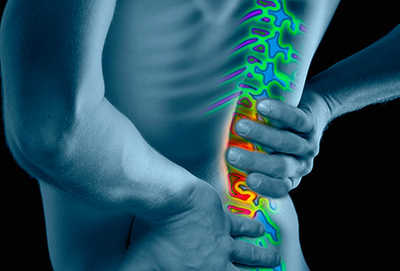
A 38-year-old active duty male presents with sudden worsening of his chronic low back pain after trying to change a vehicle tire single-handedly while you are out in the field. He states that his back pain is accompanied by severe shooting pains going down his left leg to his calf, and that he feels like the inside of his left thigh is numb, “almost like its asleep”, and his left foot “doesn’t seem to want to work”. His normal medication regimen of 800mg motrins and “some muscle relaxers” have not helped his pain at all, and it was so hard to walk that he had to lean on a buddy in order to be helped over to your aid station.
- What is your differential diagnosis at this time?
- Which diagnosis should be most concerning?
- What are some area-specific questions that you can ask this patient to better evaluate and triage them?
- What are some additional physical exam tests you can do to check and narrow your differential diagnosis list?
- What can we do about it before the patient is sent forward for further care?
- What is the definitive care that this patient requires?
1) What is your differential diagnosis at this time?
- mechanical low back pain
- simple muscle strain
- disc herniation
- sciatica/piriformis syndrome
- cauda equina syndrome
2) Which diagnosis should be most concerning?
Cauda equina syndrome (CES), a neurosurgical emergency consisting of a mass effect (i.e. compression from herniated disc, tumor, hematoma, or other traumatic or atraumatic mass) pushing against the inferior spinal cord. The cauda equina (“horse’s tail”) is where the spinal cord itself comes to an end, and all remaining spinal nerves (usually those after L1) are left exposed within the spinal column. These nerves are more susceptible to compression from mass effect than the levels above it for a number of reasons, including less protecting covering (specifically from Schwann cells) than spinal nerves above them, and no regionalized blood supply (i.e. no “back up” if some supply is lacking or is cut off).


3) What are some area-specific questions that you can ask the patient to better evaluate and triage them?
Again, CES is your chief concern given the area and severe onset. The primary “red flag questions” that should immediately point you toward the direction of emergency and immediate medevac are:
- Any loss of bowel/bladder control? or any bowel/bladder dysfunction? – this does not necessarily mean that a patient comes in urinating or defecating on himself (although that can happen). Most people don’t realize that like many of the organs inside your body, the bladder and urinary tract consist of muscle, and therefore any disruption of the motor nerves can cause dysfunction. Medical literature notes that most CES cases have urinary retention. At first, this can be extremely painful (imagine your bladder slowly stretching like a water balloon as it fills). However, as CES progresses, sensory nerves are compressed as well, and so painless urinary retention is especially troubling when assessing acute low back pain cases. Much of the CES literature divides CES presentations into “incomplete CES” when a patient reports altered urinary sensation, loss of desired to void, poor urinary stream, or straining, and “CES with retention” when the patient has painless retention.
- Any “saddle paresthesias”? – this is a question most have heard numerous times but may not truly understand, although its origins are quite obvious. Paresthesias, which can describe numbness, prickly, or tingling sensations, is often found in the upper inside thigh (super-medial thigh) and possibly genitals, when presenting in cases of CES. This occurs when the sensory nerve roots are being compressed by mass effect. While saddle paresthesias is the buzz word/catch phrase/exam answer, it is important to note that not all patients will present like this. The entire lower body is covered by the cauda equina, so these paresthesias are troubling if they present anywhere in the lower extremities with new-onset low back pain.


- Any lower extremity weakness? – as mentioned above, both sensory and motor nerves emerge from the cauda equina, and therefore patients may report perceived weakness with their back pain. Motor strength testing is reviewed below, but especially obvious on presentation would be weakness with foot dorsiflexion (raising the back of the foot up). This presents as “foot drop” when the patient walks in, where the patient is forced to raise their leg up slightly higher than normal because they cannot pick their foot up all the way.1 Patients also may report constant tripping or difficulty using stairs because of this weakness.
- Any fevers, night sweats, chills, unintentional weight loss? – although less likely in a military population (as opposed to civilian emergency departments), other possible causes of mass effect in the lower back include a spinal/paraspinal abscess associated with IV drug use (or other causes of infection), and the ever-present concern for tumors/cancer. Asking basic constitutional symptom questions like these can help to eliminate differential diagnoses quickly.
4) What are some additional physical exam tests you can do to check and narrow your differential diagnosis list?
The physical exam to help evaluate acute low back pain and rule out CES is basic in nature but must be thorough. This includes complete lower extremity motor strength and sensory testing throughout, looking for any weakness, deficits, or asymmetry.
Basic motor neuro testing involves the following areas to test for each lumbosacral nerve:

Basic sensory testing involves light and sharp touch testing at all of the skin levels annotated in the graphic below. Each of these areas corresponds to specific nerve roots.

In addition, sensory testing must include deep tendon reflexes (DTRs) at the knee and ankle, as well as the cremasteric reflex.
L1 – cremasteric reflex
L4 – patellar (knee) reflex
S1 – Achilles’ (ankle) reflex
Additionally, special tests can include:
- Straight leg raise – in this exam, the patient lies supine (on their back) and each leg is individually raised while in the straightened position. The test is considered positive when the patient has pain that is sciatic in nature when the leg is raised between 30 and 70 degrees, meaning that there is pain radiating down the back of the thigh/leg that is raised down to at least the back of their knee.2 This test is significantly sensitive for a disc herniation, meaning that if there is a herniated disc, you will likely capture it with this test.
- Contralateral straight leg raise – same exam, but in this one when the leg is raised, the patient has pain on the opposite side (i.e. raising a straightened left leg causes right lower extremity symptoms). This test is significantly specific, meaning that if it is positive, it is likely to be due to a herniated disc. Therefore the straight leg raise and contralateral straight leg raise are very complimentary in nature.
- Digital rectal exam for rectal tone – this assesses for loss of motor strength in the anal sphincter muscles, which can be caused by compression in CES.
5) What can we do about it before the patient is sent forward for further care?
In a Battalion Aid Station or other field/austere environment scenario, there is very little outside getting IV access and pain control medications that can be done for this patient. There are some case reports of using IV corticosteroids (i.e. 125mg methylprednisolone or 10mg dexamethasone) to help with inflammation as bridging care, but this is not definitive care and should not delay medevac to a higher medical element for definitive care. Any medication beyond pain control measures should be done in consultation with a higher element.
6) What is the definitive care that this patient requires?
ANY patient presenting with acute or worsening low back pain accompanied by any positive red flag questions above should be immediately seen by a higher level provider (i.e. PA, MD/DO) and likely will be sent on to for definitive imaging en route to a neurosurgical consult. Imaging will be either magnetic resonance imaging (MRI) or computed tomography (CT) scan, depending on the facility and its resources, although MRI is superior in visualizing the discs and nerves. As is evidenced in the images below, in the case of severe disc herniation and CES, the neat and consistent spinal column is disrupted by a bulging disc, and in severe cases is clearly visible.


Conclusion
Regardless, any suspected CES should be seen by a qualified surgeon (preferably a neurosurgeon) ASAP. The ultimate treatment is for emergent surgical decompression via discectomy (trimming/removing part or all of the offending disc, or other mass). Studies have been small and far between, but several have noted that the viability of these nerves can decrease rapidly within hours, and that the delay of definitive care from surgery can result in extended recovery or permanent neurological or motor disabilities.
The views expressed in this article are those of the author and do not reflect the official policy or position of the Department of the Army, the Department of Defense, or the US Government.
For additional reading:
Douraiswami B, Muthuswamy K, Naidu DK, THanigai S, Anand V. Indeterminate cauda equina syndrome: a case report. J Clin Orthop Trauma. 2016 Jan-Mar;7(1):50-54.
Fraser S, Roberts L, Murphy E. Cauda equina syndrome: a literature review of its definition and clinical presentation. Arch Phys Med Rehabil. 2009 Nov;90 (11):1964-8.
Ho ED. A case study of cauda equina syndrome. Permanente Journ. 2003;7(4):13-17.
Small SA, Perron AD, Brady WJ. Orthopedic pitfalls: cauda equina syndrome. Am J Emerg Med. 2005 Mar;23(2):159-63.
Chau AMT, Xu LL, Pelzer NR, Gragnaniello C. Timing of surgical intervention in cauda equina syndrome: a systemic critical review. World Neurosurg. 2014 Mar-Apr;81 (3-4):640-50.
Gleave J, MacFarlane R. Cauda equina syndrome: what is the relationship between timing of surgery and outcome? Br J Neurosurg. 2002;16:325-28.
Ahn UM, Ahn NU, Buchowski JM, Garrett ES, Sieber AN, Kostuik JP. Cauda euqina syndrome secondary to lumbar disc herniation: a meta-analysis of surgical outcomes. Spine 2000 Jun 15;25(12):1515-22.

Leave a comment